


Precise removal of benign breast lumps while preserving natural breast contour, volume, and aesthetic integrity.
Fibroadenomas are benign breast masses commonly seen in younger women.
When removal is indicated, surgery can both provide diagnostic certainty and preserve the surrounding breast architecture through careful access planning and tissue handling.
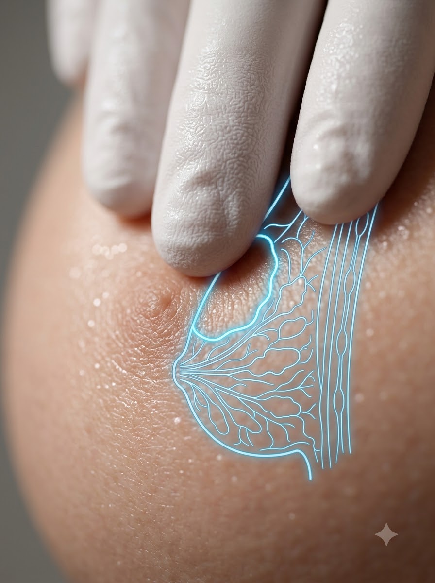
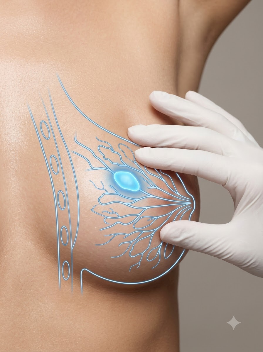
Small biopsy-proven fibroadenomas can often be monitored safely.
Symptomatic, growing, or anxiety-provoking lesions may be better managed with definitive excision.
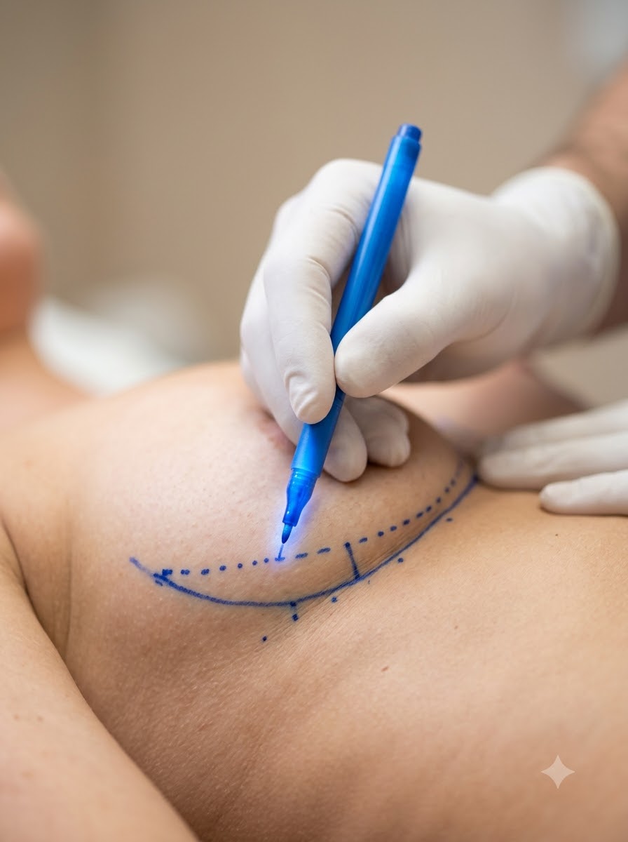

Purely functional removal can leave visible scars or contour defects.
Our approach emphasizes hidden incision placement when possible and internal reshaping so the breast maintains a smooth contour after mass removal.
"For benign breast lump surgery, how you remove the mass matters just as much as removing it."
— Clinical Director
Useful when access through the areolar border allows mass removal with a concealed scar.
Useful for lower breast lesions where the incision can hide in the fold.
Breast tissue is reshaped internally to reduce risk of contour depression after excision.
Most carefully planned benign lump excisions preserve key ducts and function, though risk depends on lesion location and extent.
Yes, removed tissue is sent for formal pathology confirmation.
Many localized excisions can be performed under local anesthesia with sedation, depending on size and location.
Recent ultrasound, mammography, or biopsy information helps guide precise planning.
Temporary avoidance helps reduce bruising and improve operative visibility.
A supportive non-underwire bra improves early post-operative comfort.
Mild soreness, swelling, and bra support are expected; arm strain should be minimized.
Return to desk work is common while avoiding upper-body impact or bouncing activity.
Contour settles, firmness softens, and activity expands as healing progresses.
The first thing most women do after finding a breast lump is reach for the worst case explanation. So let me start with the anatomy instead, because it explains why the fear is...
7 Min ReadA twenty four year old patient once sat across from Dr. Gandhi and asked the question almost every young woman asks the moment a breast lump is confirmed benign: "If it's not ca...
6 Min ReadMost young women I see for a breast lump have already spent a few sleepless nights on the internet, and by the time we meet, fear has usually outpaced facts. A fibroadenoma is a...
6 Min ReadSchedule a private consultation at our Nigdi clinic to explore your surgical options with absolute discretion and expert guidance.